Leg Bone Diagram Labeled - Leg Picture Image On Medicinenet Com : The pubis, ischium, and ilium together constitute the pelvis while the thigh bone is the femur.
byAdmin•
0
Leg Bone Diagram Labeled - Leg Picture Image On Medicinenet Com : The pubis, ischium, and ilium together constitute the pelvis while the thigh bone is the femur.. Health diagram bone skeleton leg knee science anchor chart human human body. The thigh is that portion of the lower limb located between the hip joint and knee joint.the leg is specifically the region between the knee joint and the ankle joint.distal to the ankle is the foot.the lower limb contains 30 bones. Master leg and knee anatomy using our topic page. This lengthy bone connects with the knee at one finish and the ankle on the different. The knee joint, you need a perfectly labeled diagram of the knee.
The pubis, ischium, and ilium together constitute the pelvis while the thigh bone is the femur. The blood supply to and/or from the navicular bone is disrupted. Beside that, we also come with more related ideas as follows free printable human anatomy coloring pages, lower leg muscle diagram blank and lower limb bones unlabeled. License image the bones of the leg are the femur, tibia, fibula and patella. Registered nurse, free care plans, free nclex review, nurse salary, and much more.
Skeletal System 1 The Anatomy And Physiology Of Bones Nursing Times from cdn.ps.emap.com Knee, leg, and foot (overview) how many times have a layman's language and anatomy ever matched? The hip itself is a ball and socket joint, much like the shoulder.the structures necessary to create this joint are the socket, the joint capsule, muscle, ligaments, and the neck. The thigh is that portion of the lower limb located between the hip joint and knee joint.the leg is specifically the region between the knee joint and the ankle joint.distal to the ankle is the foot.the lower limb contains 30 bones. Connective tissue is in blue. At the distal end of the femur, two rounded condyles meet the tibia and fibula bones of the lower leg to form the knee joint. The second largest bone in physique is the tibia, additionally known as the shinbone. The knee joint is the largest joint in the body and is primarily a hinge joint, although some sliding and rotation occur. Inside of a female human body bones.
There are three hamstring muscles, all of them originating at the ischial tuberosity (the bones you sit on):
These are the femur, patella, tibia, fibula, tarsal bones, metatarsal bones, and phalanges (see. Beside that, we also come with more related ideas as follows free printable human anatomy coloring pages, lower leg muscle diagram blank and lower limb bones unlabeled. Are you up to the challenge? Master leg and knee anatomy using our topic page. Color the bones of the hand human skull anatomy, anatomy bones, anatomy these pictures of this page are about:blank foot bone diagram. The femur, or thighbone, is the longest and largest bone in the human body. (there are four types of bone: Tibia and fibula bone anatomy with diagram picture and free quiz. Learn vocabulary, terms, and more with flashcards, games, and other study tools. Our goal is that these leg anatomy worksheets pictures gallery can be a direction for you, bring you more references and also make you have a great day. Health diagram bone skeleton leg knee science anchor chart human human body. Degenerative disease, similar to arthritis. Registered nurse, free care plans, free nclex review, nurse salary, and much more.
The femur is the largest bone in the body and the only bone of the thigh. It is the largest bone in the body and is the only bone in the upper leg. Our goal is that these leg anatomy worksheets pictures gallery can be a direction for you, bring you more references and also make you have a great day. Tibia and fibula bone anatomy with diagram picture and free quiz. Labeled diagram of the bones and ligaments of the foot.
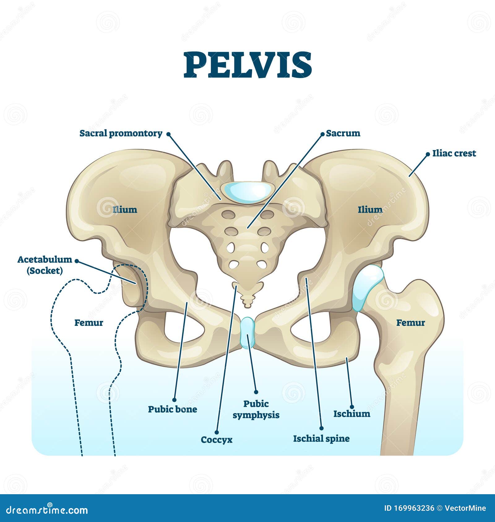
Pelvis Anatomical Skeleton Structure Labeled Vector Illustration Diagram Stock Vector Illustration Of Ischium Graphic 169963236 from thumbs.dreamstime.com Bone diagram forehead (frontal bone) nose bones (nasals) cheek bone (zygoma) upper jaw (maxilla) lower jaw (mandible) breast bone (sternum) upper arm bone (humerus) lower arm bone (ulna) thigh bone (femur) collar bone (clavicle) toe bones (phalanges) ankle bones (tarsals) kneecap (patella) shin bone Each leg is made up of four bones. Lower leg muscle diagram blank leg muscles anatomy, gross anatomy.related: The bones of the hip include the femur, the ilium, the ischium, and the pubis. However, the definition in human anatomy refers only to the section of the lower limb. Benjamin ma, md, professor, chief, sports medicine and shoulder service, ucsf department of orthopaedic surgery, san francisco, ca. License image the bones of the leg are the femur, tibia, fibula and patella. Master leg and knee anatomy using our topic page.
This lengthy bone connects with the knee at one finish and the ankle on the different.
It is likely that abnormal biomechanical stresses are the basis for the disease. The thigh bone, or femur, is the large upper leg bone that connects the lower leg bones (knee joint) to the pelvic bone (hip joint). Knee, leg, and foot (overview) how many times have a layman's language and anatomy ever matched? This page is about leg bones diagram,contains aluminium plant safety: Like the upper limb, the lower limb is divided into three regions. Inflammation of navicular bone and/or bursa. The bones of the hip include the femur, the ilium, the ischium, and the pubis. The hip joint gives the leg an incredible range of motion while still providing support to the body's weight. To understand one of the most complex joints of our body i.e. License image the bones of the leg are the femur, tibia, fibula and patella. Related posts of bones leg diagram picture bone structure horse hind leg. This will help you to understand the mechanism as well as the working. Are you up to the challenge?
The knee joint, you need a perfectly labeled diagram of the knee. (there are four types of bone: The femur is the largest bone in the body and the only bone of the thigh. Knee, leg, and foot (overview) how many times have a layman's language and anatomy ever matched? Diagram and names of leg bones, diagram of foot and leg bones, diagram of leg bones, diagram of lower leg bones, diagram of the bones in your leg, bone, diagram and.
Label The Lower Leg Diagram Quizlet from o.quizlet.com The knee joint is the largest joint in the body and is primarily a hinge joint, although some sliding and rotation occur. It is the largest bone in the body and is the only bone in the upper leg. Color the bones of the hand human skull anatomy, anatomy bones, anatomy these pictures of this page are about:blank foot bone diagram. Are you up to the challenge? (there are four types of bone: The bones of the leg are the femur, tibia, fibula and patella.the foot bones shown in this diagram are the talus, navicular, cuneiform, cuboid, metatarsals and calcaneus. Each leg is made up of four bones. This muscle runs along the outside of the back of your thigh and attaches to the top of the fibula (the smaller of the two bones of your lower leg).
The foot bones shown in this diagram one of the beloved.
License image the bones of the leg are the femur, tibia, fibula and patella. This will help you to understand the mechanism as well as the working. Labeled human leg bones created for use in leg bone. At the distal end of the femur, two rounded condyles meet the tibia and fibula bones of the lower leg to form the knee joint. Also called the thigh bone, this is the longest bone in the body.it. This image is an edited version of this image that was created by user:ladyofhats (mariana ruiz villarreal). These are the femur, patella, tibia, fibula, tarsal bones, metatarsal bones, and phalanges (see. The hip itself is a ball and socket joint, much like the shoulder.the structures necessary to create this joint are the socket, the joint capsule, muscle, ligaments, and the neck. The blood supply to and/or from the navicular bone is disrupted. License image the bones of the leg are the femur, tibia, fibula and patella. Posted on april 18, 2019april 18, 2019. Long bones are found in the arms (humerus, ulna, radius) and legs (femur, tibia, fibula), as well as in. The upper leg is often called the thigh.
There are three hamstring muscles, all of them originating at the ischial tuberosity (the bones you sit on): leg bone diagram. The hip joint gives the leg an incredible range of motion while still providing support to the body's weight.